

List of the Quiz
| About Author | |
| Authors | Youe Ree Kim |
| Institution | Wonkwang University Hospital |
| sweetynn@naver.com | |
| About Case | |||
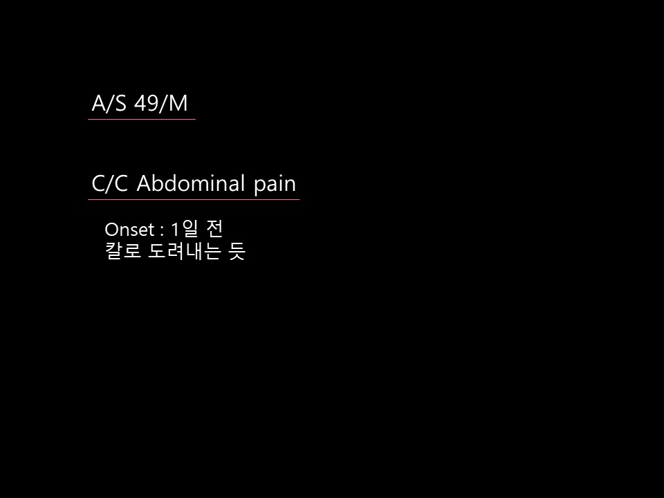
| Chief complaint or Title |
Abdominal pain | ||
| Age | 49 | Sex | M |
| Difficulty | For resident, For specialist | ||
| Modality | US, CT | ||
| Case Figures | |||||||||
|
|||||||||
| Questions | ||
| What is the cause of intussusception? |
| Answer | |||
| Diagnosis | Colonic anisakiasis (Infectious colitis) | ||
| Comments | The patient was diagnosed with colonic anisakiasis on endoscopy. On serological examination, except for mild CRP elevation (25.1), complete blood counts including white blood cells were normal, and there was no peripheral eosinophilia. Bowel wall thickening and pericolic haziness improved on CT image after albendazole treatment.
Gastrointestinal anisakiasis is a rare parasitic disease in human beings and it is caused by penetration of a nematode larva belonging to the family Anisakiadae. It is most commonly associated with the stomach wall and small intestines. Colonic anisakiasis is rare. The most reasonable explanation for the rarity of this colonic infection is that the colon is too far for the orally ingested larvae to attain the colon. Adult intussusception caused by anisakiasis is extremely rare. Clinical presentations - Acute abdominal pain of varying severity, nausea, vomiting, urticaria, chills and diarrhea - Within a few hours after ingestion of raw fish - Leukocytosis : common laboratory finding - Peripheral eosinophilia : frequently gastric form, uncommon finding in cases of intestinal anisakiasis CT findings - Long segment of circumferential and symmetric thickening of the bowel wall - Luminal narrowing and diffuse mucosal enhancement (target sign) - Ascite -> Colonic anisakiasis may simulate a tumor of the colon, since this infection provokes edema, an acute phlegmonous reaction, or granuloma formations around the larvae in the submucosa of the intestinal wall, which results in a mass effect
|
||
| References | Korean J Radiol 2008;9:S56-60 J Korean Soc Radiol 2011;64:167-171 J Nippon Med Sch 2006;73:169-174 World J Gastroenterol 2010;16:1804-1807 |
||
| Keywords | Infection, Parasite, Anisakiasis, Colon | ||
| Attachment File | - | ||


Number of Applicants : 53
| Correct Answer | 5 |
| - | 변성환 |
| 삼성서울병원 | 신재승 |
| 고려대구로병원 | 이다경 |
| 중앙보훈병원 | 임정수 |
| 중앙보훈병원 | 조상현 |