
Quiz of the Week
| About Author | |
| Authors | Shin Yong Moon |
| Institution | Asan Medical Center |
| cjimage | |
| About Case | |||
| Chief complaint or Title |
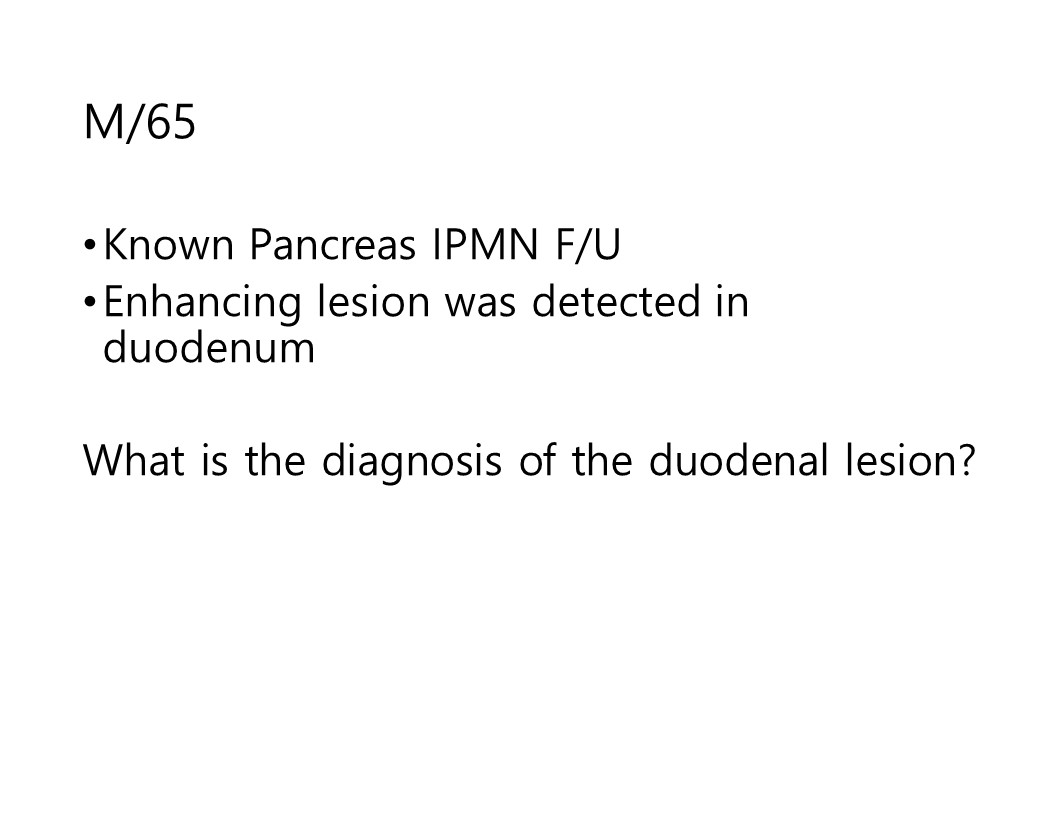
incidental duodenum mass | ||
| Age | 65 | Sex | M |
| Difficulty | For resident, For specialist | ||
| Modality | CT, MR | ||
| Case Figures | |||||||||
|
|||||||||
| Questions | ||
| What is your diagnosis |
| Answer | |||||||||||||
| Diagnosis | 3 | ||||||||||||
| Comments | Clinical course ; Patient had IPMN, and 1cm lesion in duodenum Duodenum 3rd/4th
portion enhancement 1cm nodular lesion R11; no change of size, T2 intermediate SI -
Diagnosed at subepithelial
tumor such as GIST, neuroendocrine tumor -
PPPD was done and duodenal
lesion was revealed as Dilated submucosal vein, -
consistent with venous malformation,
duodenum. Vascular malformation 1.
Low-flow vascular malformations - Capillary malformation - Venous malformation - Lymphatic malformation 2.
High-flow vascular
malformations - Arterial malformation - Arteriovenous malformation - Arteriovenous fistula 3.
Combined vascular malformation
(combination of above lesion types) The most common site of AVM is the cecum
and right colon (78%), followed by the jejunum (10.5%), whereas only 0.9% of
all AVMs are found in the pancreas. The cause of pancreatic AVM is thought to
be congenital in 90% of cases, and 10% to 30% of pancreatic AVM are associated
with Osler-Weber-Rendu disease. Several cases of acquired pancreatic AVM
occurred because of pancreatitis, trauma, and tumors. After enhancement, early contrast filling
of the enlarged portal venous system was seen in the arterial phase. In
addition, demonstration of enhancement of the lesion, commensurate with the
aorta, on contrast enhanced multiphasic imaging is helpful in diagnosing
pancreatic AVM Characteristic signal void on T1- and
T2-weighted imaging could provide the diagnostic clue for AVM. This signal void is a characteristic of
rapid blood flow. However, slow flow lesion shows atypical
signal in MRI. MR features of vascular malformation.
|
||||||||||||
| References | Usman Bashir, et al. Magnetic Resonance (MR) Imaging of Vascular Malformations. Pol J Radiol. 2017; 82: 731R11;741. Borahm Lee, et al. Pancreatic Arteriovenous Malformation as an Unusual Cause of Chronic Gastrointestinal Bleeding in a Patient with Early Gastric Cancer:Multimodality Imaging Spectrum with Pathologic Correlation. iMRI 2015;19:241-247 | ||||||||||||
| Keywords | duodenum, vascular malformation | ||||||||||||
| Attachment File | - | ||||||||||||

Number of Applicants : 58
| Correct Answer | 20 |
| Shiga University of Medical Science | Akitoshi Inoue |
| University of Tokyo hospital | Jun Kanzawa |
| Fukuoka University | Keisuke Sato |
| Shiga University of Medical Science | Ryo Uemura |
| Osaka Metropolitan University hospital | Tatsushi Oura |
| University of Tokyo | Toshihiro Furuta |
| 용인세브란스병원 | 강예슬 |
| 서울아산병원 | 구보연 |
| 서울아산병원 | 김경원 |
| 한림대성심병원 | 김민정 |
| 순천향대부천병원 | 박혜주 |
| - | 변성환 |
| 삼성서울병원 | 신재승 |
| 경희의료원 | 이용대 |
| 보라매병원 | 장시원 |
| 서울특별시 서울의료원 | 정하택 |
| 중앙보훈병원 | 조상현 |
| 경희의료원 | 조현철 |
| 서울대학교병원 | 홍성호 |
| 서울의료원 | 홍혁기 |