
Quiz of the Week
| About Author | |
| Authors | Soo Jin Kim |
| Institution | NCC |
| sjkim@ncc.re.kr | |
| About Case | |||
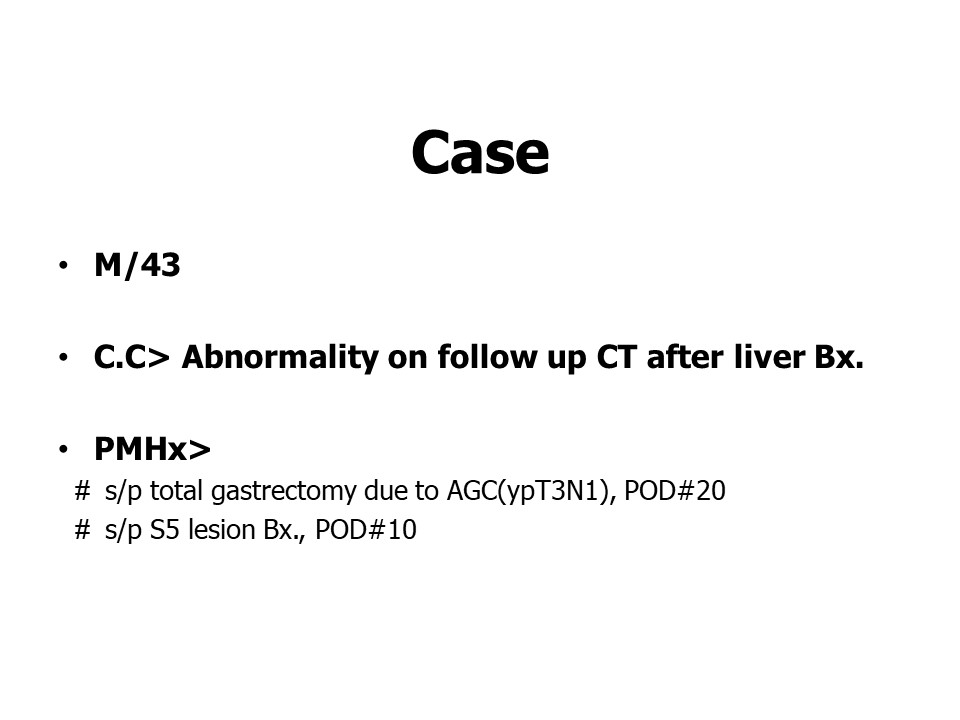
| Chief complaint or Title |
Abnormality on follow up CT after liver biopsy | ||
| Age | 43 | Sex | M |
| Difficulty | For resident, For specialist | ||
| Modality | CT, Angiography | ||
| Case Figures | |||||||||
|
|||||||||
| Questions | ||
| What's your diagnosis? |
| Answer | |||
| Diagnosis | Recurrent bleeding from pseudoaneurysm of Lt. inferior phrenic artery. | ||
| Comments |
Precontrast CT shows high attenuated fluid-debris level in the pelvis along dependent area suggesting hemoperitoneum and acute/subacute state of recent bleeding.
It is always challengable but doable to detect the orgin of bleeding. When I found out a tiny saccular abnormal structure corresponding to Lt. inf. phrenic artery, the main operator who conducted total gastrectomy also suspected iatrogenic injury to that vessel by electrocautry during the operation. Diagnostic and therapeutic angiography was performed. However, actually the embolization for that tiny pseudoaneurysm was technically failed due to very stiff angulation of celiac os. Then, interventional radiologist just performed the embolization at S6 biopsy site with suspicion of woozing and possibility of delayed bleeding from the biopsy site also. Pathology report for S5/6 biopsy lesion was hemangioma, clinically suspected as early liver mets.
Two weeks later, when patient showed hypotension, CT angiography definitely revealed increased size of a saccular pseudoaneurysm of Lt. inf. phrenic artery especially it was well demonstrated on MRP and 3D reconstruction images. After multiple trials with every different interventional devices, finally the embolization for LIPA pseudoaneurysm had succeeded, the saccular filling of contrast media was disappeared on postembolization spot image.
|
||
| References | 1) Pasha SF, et al. Splanchnic artery aneurysms. Mayo Clin Proc. 2007;82(4): 472-479 2) Shanley CJ, et al. Uncommon splanchnic artery aneurysms: Pancreaticoduodenal, gastroduodenal, superior mesenteric, inferior mesenteric, and colic. Ann Vasc Surg. 1996;10:506-515 3) Panayiotopoulos YP. et al. aneurysms of the visceral and renal arteries. Ann R Coll Surg Engl. 1996;78:412-419 | ||
| Keywords | pseudoaneurysm, inferior phrenic artery, intraoperative trauma | ||
| Attachment File | - | ||


Number of Applicants : 35
| Correct Answer | 27 |
| 의정부성모병원 | 이수림 |
| Mayo Clinic | Akitoshi Inoue |
| The University of Tokyo Hospital | Toshihiro Furuta |
| 서울아산병원 | 김경원 |
| 서울아산병원 | 김동욱 |
| 한림대성심병원 | 김민정 |
| 세브란스 | 김승섭 |
| 서울의료원 | 김여은 |
| 세브란스병원 | 김연윤 |
| 중앙보훈병원 | 김윤이 |
| 중앙보훈병원 | 김주희 |
| 서울아산병원 | 김지은 |
| 중앙보훈병원 | 노지훈 |
| 군의관 | 문경일 |
| 해운대백병원 | 손정희 |
| 서울아산병원 | 신용문 |
| 세브란스병원 | 신재승 |
| 고대구로병원 | 이석영 |
| 고대구로병원 | 이유경 |
| 로컬 | 이현규 |
| 중앙보훈병원 | 장성원 |
| 보라매병원 | 장시원 |
| 고대안산병원 | 최벽경 |
| 고대구로병원 | 최재웅 |
| 고대안산병원 | 하모인 |
| 서울대병원 | 한승철 |
| 서울의료원 | 홍혁기 |